Medical Imaging
Our interests are in developing diagnostic applications and strategies for the acquisition and visualization of 2 or 3-dimensional medical imaging data. Recently, we investigate on the diagnosis of the optic nerve related disease using 3-dimensional functional image analysis technique.
Ongoing Project
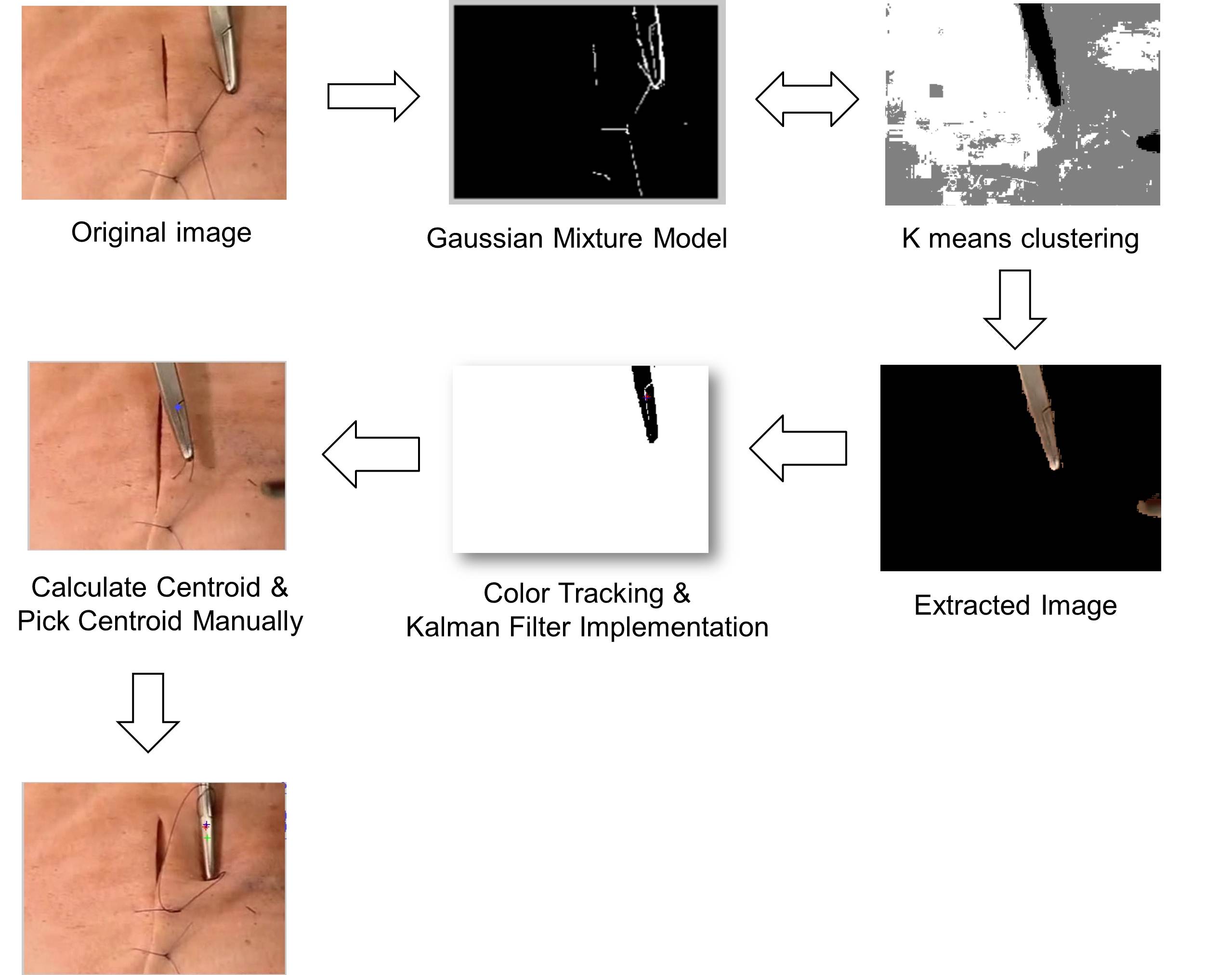
Image Processing in Robot-assisted Laparoscopic surgery
- Real-time Segmentation of Surgical Instruments
As automation and use of robotics, such as surgical navigation and medical robot control, plays an ever increasing role in telemedicine field today
To precisely control the robot, accurate segmentation of surgical tool regions in laparoscopic images is necessary.
Based on this environment, fast segmentation of tool locations with diminishing outliers due to time-varying lighting conditions and background noise needs to be implemented.
Retinal Image Analysis
- Quantification of Retinal Nerve Fiber Layer Atrophy
Glaucoma is a group of diseases caused by the damage of the optic nerve, resulting in visual field loss and blindness. Early stage diagnosis and treatment is important, because the loss of vision is irrevocable. Retinal nerve fiber atrophy (RNFL) is a defect caused by death of nerve cell. Measuring the atrophy and its progression is vital to early detection and management of glaucoma. For quantitative analysis, several methods were suggested, but they are not completely quantitative and automatic. In fundus photograph, the region with RNFL atrophy shows lower intensity compared to normal RNFL. So we developmed the novel method based on intensity profile of the pixels on the circle around the optic nerve head using image processing and learning techniques.
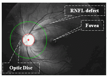
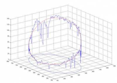
<Retinal fundas image & Intensity profile of RNFL>
Stereo Imaging & 3D Vision
- 3D Reconstruction of Optic Nerve Head(ONH) from Stereo Disc Photograph (SDP)
Galucoma is a disorder in which the pressure in the eyeball increase, damaging the optic nerve and causing a loss of vision. Early detection of ONH deformation is important in the treatment of Galucoma. The 3D reconstruction from SDP enable to detect and standarize ONH deformation with low-cost.
Stereo disc photograph was analyzed and reconstructed as 3 dimensional contour image to evaluate the status of the optic nerve head for the early detection of glaucoma and the evaluation of the efficacy of treatment. Stepwise preprocessing was introduced to detect the edge of the optic nerve head and retinal vessels and reduce noises. Paired images were registered by power cepstrum method and zero-mean normalized cross-correlation. After Gaussian blurring, median filter application and disparity pair searching, depth information in the 3 dimensionally reconstructed image was calculated by the simple triangulation formula. Calculated depth maps were smoothed through cubic B-spline interpolation and retinal vessels were visualized more clearly by adding reference image. Resulted 3 dimensional contour image showed optic cups, retinal vessels and the notching of the neural rim of the optic disc clearly and intuitively, helping physicians in understanding and interpreting the stereo disc photograph.
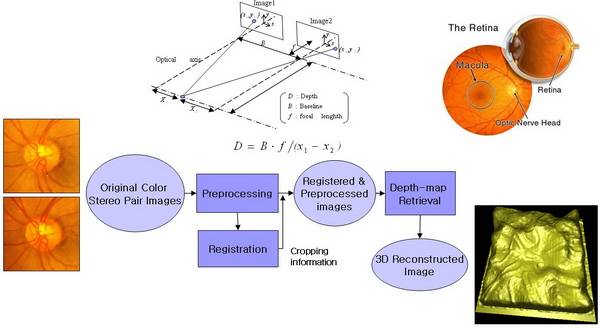
<3-dimensional reconstruction of ONH using stereo disk photograph>
Elapsed Project
Acne vulgaris Image Analysis
- Detection of Acnes in whole face region
Acne vulgaris is a chronic and common inflammatory disease of the pilosebaceous unit.

For continuous and effective skin care, computerized acne photographic systems are commonly used. But the systems do not provide screening for the entire face. To overcome this drawback, we devised a face acne capturing system which is composed of a DSLR camera, illuminations, and a rotating mount. Patient’s chin was placed on a chin orthotic. The face was viewed and captured in real time using PC connected to the camera. The square-shape frame CCFL provides uniform illumination to the face. Semi-circular rotatable mount enables the camera to take pictures of curved surfaces from the entire face.
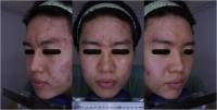
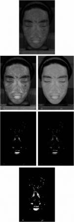
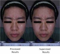
Acquired images have shown that inflammatory acne regions were properly detected after color space conversion, intensity adjust, morphological operations and binarization methods.
By comparing the result with the manual counting by a dermatologist, the proposed system achieves reliability in analyzing acne.
Automatic Identification of Human Parasitic Eggs
In order to automate routine fecal examination for parasitic diseases, the authors propose in this study a computer processing algorithm using digital image processing techniques and an artificial neural network (ANN) classifier The morphometric characteristics of eggs of human parasites in fecal specimens were extracted from microscopic images through digital image processing. An ANN then identified the parasite species based on those characteristics. The authors selected four morphometric features based on three morphological characteristics representing shape, shell smoothness, and size. A total of 82 microscopic images containing seven common human helminth eggs were used. The first stage (ANN-1) of the proposed ANN classification system isolated eggs from confusing artifacts. The second stage (ANN-2) classified eggs by species. The performance of ANN was evaluated by the tenfold cross-validation method to obviate the dependency on the selection of training samples. Cross-validation results showed 86.1% average correct classification ratio for ANN-1 and 90.3% for ANN-2 with small variances of 46.0 and 39.0, respectively. The algorithm developed will be an essential part of a completely automated fecal examination system
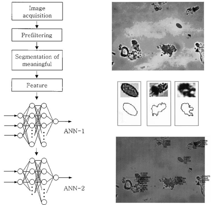
<Whole procedure of automatic Identification of Human Parasitic Eggs>
Computer-aided Diagnosis for Breast Cancer
A computer-aided diagnosis (CAD) algorithm identifying breast nodule malignancy using multiple ultrasonography (US) features and artificial neural network (ANN) classifier was developed from a database of 584 histologically confirmed cases containing 300 benign and 284 malignant breast nodules. The features determining whether a breast nodule is benign or malignant were extracted from US images through digital image processing with a relatively simple segmentation algorithm applied to the manually preselected region of interest. An ANN then distinguished malignant nodules in US images based on five morphological features representing the shape, edge characteristics, and darkness of a nodule. The structure of ANN was selected using k-fold cross-validation method with k=10. The ANN trained with randomly selected half of breast nodule images showed the normalized area under the receiver operating characteristic curve of 0.95. With the trained ANN, 53.3% of biopsies on benign nodules can be avoided with 99.3% sensitivity. Performance of the developed classifier was reexamined with new US mass images in the generalized patient population of total 266 (167 benign and 99 malignant) cases. The developed CAD algorithm has the potential to increase the specificity of US for characterization of breast lesions.
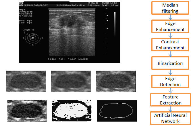
<Example of ultrasound breast nodule image & Whole procedure of analysis>
Ultrasound Measurement of Residual Urine Volume According to Bladder Shape
To suggest a novel algorithm based on individually determined proportionality constants to improve the accuracy of noninvasively calculated residual urine volume using ultrasound bladder images. The residual urine volume of 116 subjects was calculated from ultrasound images obtained using a conventional two-dimensional ultrasound device at two institutions. The proposed method uses individually determined proportionality constants for specific bladder shapes. The corresponding true residual volumes were determined by measuring the catheterized volume. The accuracy of our method was confirmed by comparing the mean and standard deviation of the fractional absolute error (FAE) versus true volume with values obtained using other methods, which rely on fixed constants. The calculated residual bladder volume of 116 patients correlated significantly (r = 0.94, P <0.001) and the differences from the true values were not statistically significant (P = 0.42). The mean FAE was 0.17 ± 0.10. Subgroups divided by sex, postvoid residual urine volume (over and under 150 mL), and institution produced different results (mean FAE for men 0.15 ± 0.11 and for women 0.18 ± 0.12; mean FAE for greater than 150 mL, 0.16 ± 0.11 and for less than 150 mL, 0.17 ± 0.14; mean FAE for Seoul National University Hospital 0.15 ± 0.10 and for Seoul National University Bundang Hospital 0.18 ± 0.18). Our method provided better accuracy than other known formulas using a conventional two-dimensional ultrasound scanner. The latter multiply the product of the sagittal height and depth and transverse width by a fixed coefficient.
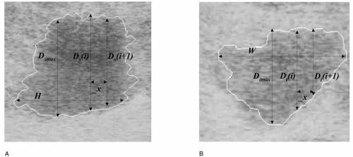
<Examples of ultrasound images and detected boundaries of residual urine in (A) sagittal and (B) transverse planes, in which the three dimensions (H, D, and W) and two coefficients (α and β) were automatically determined>

