Bio-MEMS
In recent issues on biotechnology(BT) area, the need for creating and applying a microscale systems suggests the need to implement all the factors lighter, thinner, shorter and smaller. Research on these microscale systems is highly active due to its technical and economical benefits and prospects, namely:
(1) Reagents used for molecular biology analysis are mostly high priced and therefore a means to minimize the reagents' quantity is necessary.
(2) The amount of samples used for analysis is becoming smaller and smaller, so a method to allow the usage of small amounts of samples is also needed.
Regarding technological benefits, new techniques such as follows have been developed and their medical and biological application is being implemented. Photolithography, a method used for fabricating integrated circuits(IC) with semiconductors, is used. This system that uses photolithography dor fabrication is called Micro Electro Mechanical Systems(MEMS). Therefore, MEMS refers to microscale systems or microscale precision machines, and is also called microsystems, micromachines, and micromechatronics. These MEMS technology allows fabrication of tiny parts simultaneousely and can embed electrical components during the fabrication process, and therefore it has the advantage of scaling down the size of the whole system even greater.
Components manufactured my MEMS technologies are usually several ㎛ in size, and the whole system is several of millimeter order, but it is a complete system by itself. In applications for medical and biological fields, neurochip, cell chip, DNA chip, biochips such as lab-on-a-chip, microelectrodes for biosensors, microneedles for drug delivery or painless bodily fluid retreaval are some of its application examples.
MEMS technology application into medical and biological areas have given rise to a new fied of study called BioMEMS, and microscale surgical tools, artificial organs, various diagnosis tools, drug delivery tools are explored in implementing the MEMS technology in these fields. Two technologies used for BioMEMS in these fields are polymer MEMS that uses plastic materials than silicon, and microfluidics that uses ㎛-sized fluid channels. In figure 5, there are examples of technological research development in these BioMEMS techniques.
In addition to these applications, interests in nanotechnology, which deals in manufacturing and using carbon nanotubes and other nanoscale structures are growing fast, and research to develop medical treatment tools and to proceed with various diagnosis using Nano Electro Mechanical Systems(NEMS) is in progress. So far there are many technical hardships, both theoretically and technically, in making full use of this technology, but once the biosensors of nanometer scale is combined with microfluidics, micro total analysis(μTAS) to its fullest is possible using a single small biochip for all types of blood analysis. Also, this allows the realization of single cell workbench, which can distribute single cells to the desired location, stimulate the interior and exterior cellular fluids, anlyze secretion from the cell, inject, extract, and analyze DNAs, RNAs, proteins and other genetic materials. This technology will advance into microsystems that flows within human blood vessels and detect cancers at early levels or suppress their activity or hopefully exterminate them.
Ongoing Project
- DNA hybridization using the Microfluidic Chip - Jongmin Noh
- Blood Electrolyte and Gas sensor - Jongmin Noh
- High Throughput Decoding System for Multiplex Suspension Array Analysis - Saram, Lee
- High speed CTC(Circulating Tumor Cell) Screening System Using Microfludic Chip - Kwang Bok Kim, Hyoungseon Choi
- Gold Nano-Particle Preconcentration for SERS Analysis On Microchip System - Kwang Bok Kim
- A Microfluidic Chip-based Micro Flow Cytometry with Optical and Electrical method - Hyoungseon Choi
Elapsed Project (Published)
- Flow Cytometry-based Submicron-Sized Bacterial Detection System using Movable Virtual Wall - Hyoungseon Choi
- Hyoungseon Choi, Chang Su Jeon, Inseong Hwang, Juhui Ko, Saram Lee, Jaebum Choo*, Jin-Hyo Boo, Hee Chan Kim* and Taek Dong Chung*, “Flow Cytometry-based Submicron-Sized Bacterial Detection System using Movable Virtual Wall”, Lab on a Chip, 2014, 14(13), 2327-2333
- A label-free DC impedance-based microcytometer for circulating rare cancer cell counting - Hyoungseon Choi
- Hyoungseon Choi , Kwang Bok Kim , Chang Su Jeon , Inseong Hwang , Saram Lee , Hark Kyun Kim* , Hee Chan Kim* and Taek Dong Chung*, “A label-free DC impedance-based microcytometer for circulating rare cancer cell counting”, Lab on a Chip, 2013, 13, 970-977

* Microfluidic Chip based Polyelectrolyte Junction Field Effect Transistor. - Kwang bok Kim
- Kwang Bok Kim, Ji-Hyung Han, Hee Chan Kim* and Taek Dong Chung*, “Polyelectrolyte Junction Field Effect Transistor based on Microfluidic Chip”, Applied Physics Letters, 2010, 96(14), 143506
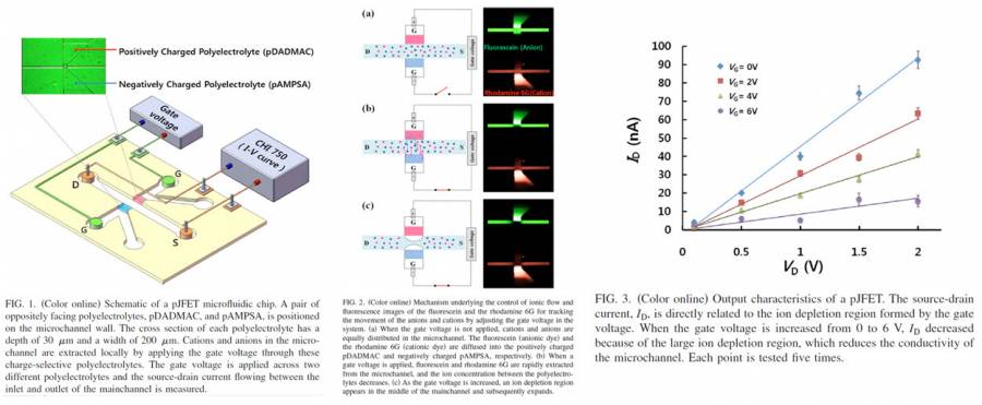
Picture 1. shows a pJFET system that operates in an aqueous medium by reliably controlling the ionic flow through oppositely charged polyelectrolyte plugs.
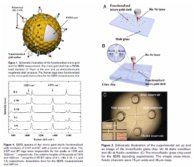
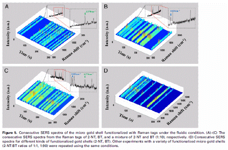
- SERS Decoding of Micro Gold Shells Moving in Microfluidic Channel - Sa Ram Lee
- Saram Lee, Segyeong Joo, Sejin Park, Soyoun Kim, Hee Chan Kim* and Taek Dong Chung*, “SERS Decoding of Micro Gold Shells Moving in Microfluidic Systems”, Electrophoresis, 2010, 31, 1-7
* Microfludics(Lab-on-a-chip)-based FACS(Fluorescence Activated Cell Sorter) System - Kee Hyun Kim
- Segyeong Joo, Kee Hyun Kim, Hee Chan Kim, and Taek Dong Chung, “A Portable Microfluidic Flow Cytometer based on Simultaneous Detection of Impedance and Fluorescence”, Biosensors & Bioelectronics, 2010, 25, 1509-1515, full text

Picture 1. shows the schematic of normal FACS machine. A Fluorescent activated cell sorting method is basically a device that
activates the dye's fluorescence using light sources. The activated fluorescence is then detected by using light sensors such as PMTs. This method uses laser and other bulky devices, and thus have the disadvantage of size and various hardships. Therefore, in our group, we are implementing various ways to improve the FACS machine by applying various methods in recognizing particles targeted with flourescent dyes. (Picture taken from http://csm.colostate-pueblo.edu, http://www.beckmancoulter.com, www.sysmex.co.jp/en/labscience/ )
* Particle Count on a Microfluidic Chip Using Multi Polyelectrolytic Salt Bridges. - Kwang bok Kim
- Kwang Bok Kim, Honggu Chun, Hee Chan Kim*, and Taek Dong Chung*, “Red Blood Cell Quantification Microfluidic Chip Using Polyelectrolytic Gel Electrodes”, Electrophoresis, 2009, 30, 1-6, full text

Picture 1. Microfluidic glass chip for particle counter
Picture 1. shows that the number of particles(or microbeads) is measured by microfluidic chip. The two fluidic reservoirs connected to each salt bridge were connected to an RLC meter. The RLC meter signal between the salt bridges with particles passing through the detection volume of microchannel determine the size of particles. Using the Multi Polyelectrolytic salt bridges enable single microfluidic chip to measure the multiplicity size of particles.
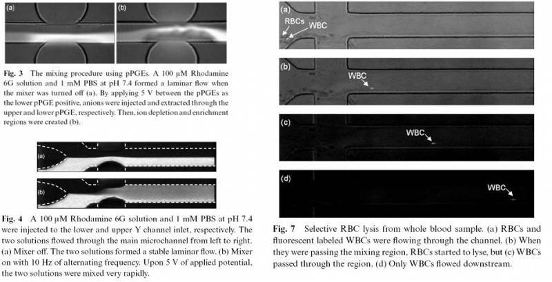
- Ultrafast active mixer using polyelectrolytic ion extractor
- Honggu Chun, Hee Chan Kim and Taek Dong Chung, Lab on a chip, 2008 Full Text
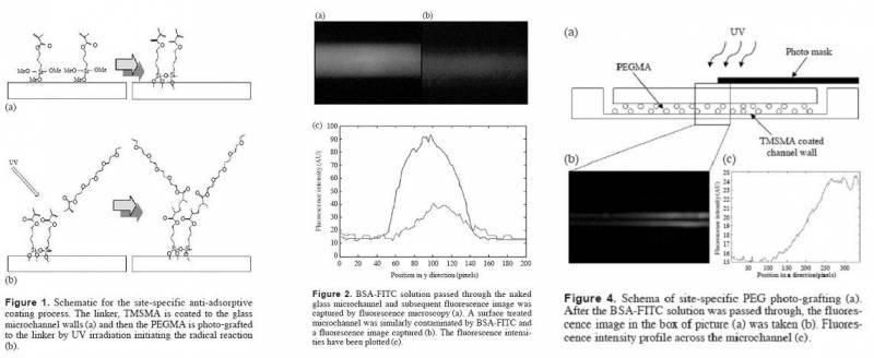
- Non Protein Adsorption Channel Coating
- Sangyun Park Hee Chan Kim+ and Taek Dong Chung+, Biochip Journal, 2007 Full Text
- A RAPID FIELD-FREE ELECTROOSMOTIC MICROPUMP INCORPORATING CHARGED MICROCHANNEL SURFACES
- Segyeong Joo, Taek Dong Chung, and Hee Chan Kim, Sensors and Actuators B, 2007 Full Text
- Integration of a Nanoporous Platinum Thin Film into a Microfluidic System for Nonenzymatic Electrochemical Glucose Sensing
- Segyeong Joo, Sejin Park, Taek Dong Chung, and Hee Chan Kim, Sensors and Analytic Science, 2007 Full Text
- //Cytometry and Velocimetry on a Microfluidic Chip Using Polyelectrolytic Salt Bridges
- Honggu Chun, Taek Dong Chung, and Hee Chan Kim / Analytical Chemistry,2005 Full Text

Equipments
Members
Related Resources
- Collaborators
T.D. Chung Group http://niel.or.kr
- Other Research Groups
S. H. Kwon Group http://binelx.snu.ac.kr/
T. S. Kim Group http://nbsl.kist.re.kr/Teams/biomems/main.htm/
J. Han Group http://www.rle.mit.edu/micronano/
S. H. Lee Group http://biomems.korea.ac.kr/
L. P. Lee Group http://biopoems.berkeley.edu/
R. Langer Group http://web.mit.edu/langerlab/
S. Bhatia Group http://lmrt.mit.edu/
P. S. Doyle Group http://web.mit.edu/doylegroup/
A. Folch Lab http://faculty.washington.edu/afolch/
A. Khademhosseini Lab http://www.tissueeng.net/lab/
S. Yang Group http://msfl.kaist.ac.kr/
K. Suh Group http://nftl.snu.ac.kr/
- Journals
Science http://www.sciencemag.org/
Nature http://www.nature.com
Nature Materials http://www.nature.com/nmat/
Nature Nanotechnology http://www.nature.com/nnano/
Nature Biotechnology http://www.nature.com/nbt/
Nature Photonics http://www.nature.com/nphoton/
Nature Methods http://www.nature.com/nmeth/
Proceedings of the National Acamedy of Sciences (PNAS) http://www.pnas.org/
Journal of the American Chemical Society (JACS) http://pubs.acs.org/journals/jacsat/index.html
Lab on a Chip http://www.rsc.org/publishing/journals/LC/
Applied Physics Letters http://apl.aip.org/ Nano Letters http://pubs.acs.org/journals/nalefd/index.html
Optics Letters http://ol.osa.org/
Optics Express http://www.opticsexpress.org/
Analytical Chemistry http://pubs.acs.org/journals/ancham/
Journal of Chromatography A http://www.elsevier.com/wps/find/journaldescription.cws_home/502688/description#description
Electrophoresis http://www3.interscience.wiley.com/journal/10008330/home?CRETRY=1&SRETRY=0
Journal of Micromechanics and Microengineering http://www.iop.org/EJ/journal/JMM
- Conferences
Micro TAS http://www.microtas2008.org/
Materials Research Society http://www.mrs.org/
IEEE MEMS http://www.mems2009.org/
Korean Bio Conference http://www.biochip.or.kr/


